|
For further MBL News and Media Information, contact the MBL Communications Office at (508) 289-7423 or e-mail us at comm@mbl.edu
FOR IMMEDIATE RELEASE: August 6, 2010
Contact: Diana Kenney, MBL, 508-289-7139; dkenney@mbl.edu
August 2010 LabBits: Science Highlights from the MBL
MBL, WOODS HOLE, MA—From cuttlefish camouflage and lamprey spinal cord regeneration to the mental map upon which memories are plotted, scientists at the Marine Biological Laboratory (MBL) are exploring some fascinating phenomena this summer.
Summer on Cape Cod is synonymous with a surge of tourists, but also a surge of visiting scientists at the MBL. For more than a century, researchers have come to the MBL each summer from around the world to immerse themselves in biological discovery. Researchers enjoy the casual, collaborative atmosphere, the access to high-tech equipment and expertise, and the escape from academic duties at their home institutions. Here is a sampling of some of the research underway this summer at the MBL’s Whitman Center for Visiting Research:
|
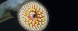
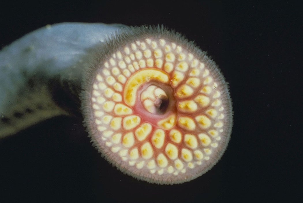
Learning from Lampreys
The super powers of an aquatic parasite, the lamprey, could provide insight into central nervous system damage and repair in humans. Eel-like in appearance, the lamprey can regenerate its spinal cord after it’s been completely severed.
“This spontaneous regeneration has been known for a long time,” says Ona Bloom of The Feinstein Institute for Medical Research. “But the molecular changes underpinning the lamprey’s ability to regenerate are not very well understood.”
Bloom and Joseph Buxbaum, of Mount Sinai School of Medicine, visiting researchers at the MBL, are spending the summer building and using molecular tools to study lamprey regeneration. Over the past year, they have collaborated with other lamprey researchers to study the lamprey transcriptome, the set of genes that are expressed in the animal at a given point in time. By studying the changes in the transcriptome over the course of spinal cord injury and regeneration, they hope to identify particular genes that are important for the process.
“The transcriptome allows us to take an unbiased approach,” Buxbaum says. “We tend to look under the lamppost, at what we know, but there could be something major that we simply don’t know about. Looking at global gene expression will ultimately lead to targeted questions about specific genes.”
Changes in the transcriptome of mammals unable to regenerate their spinal cords have already been recorded over the course of injury. “We are especially interested to know what happens at the time points when a lamprey starts to regenerate after spinal cord injury,” Bloom says.
Bloom has already been investigating the expression of specific proteins that may play a role in the immune system’s response after spinal cord injury. The gene she is focusing on codes for a protein called MIF, a molecule that promotes inflammation after injury. Inflammation contributes to the inhibition of recovery after spinal cord injury.
“There are known small molecules that can inhibit inflammation, so we can test those things in the lamprey and see what the effects are on regeneration,” Bloom says. “Inflammation is a process that’s very common in neurodegenerative diseases, so the targets we’re looking for may be applicable to other pathological settings (including multiple sclerosis and Alzheimer’s disease) in the nervous system, not just spinal cord injury.”
Bloom has confirmed that expression of MIF is induced by injury, as it is in mammals. Bloom, Buxbaum and their colleagues are identifying and investigating this and other target proteins, with the hope that their research could ultimately lead to better clinical treatments for human spinal cord injury.
“There is no effective therapy for spinal cord injury, but many patients receive a very global and powerful anti-inflammatory that can put the patient at risk for infection,” Bloom says. “Scientists are therefore searching for anti-inflammatories that are targeted to a particular molecule or kind of molecule, rather than shutting down the whole immune system.”
Bloom and Buxbaum’s inquiries into lamprey spinal cord regeneration are accelerating as they and their colleagues build up the transcriptome database. “We can take a gene and query our database, asking did it go up or down over the course of injury,” Buxbaum says. “Things that used to take a year of struggle now take 10 minutes.”
Bloom and Buxbaum recognize that the database may be useful to many researchers, including evolutionary biologists. “Our ultimate goal is to make a resource not only for our own research, but for the larger community,” Bloom says.
Bloom and Buxbaum’s summer research at the MBL is being supported by a Charles Evans Foundation Research Award. Their close collaborators at the MBL include Jennifer Morgan of University of Texas-Austin and Avis Cohen of the University of Maryland.
More information on lamprey regeneration research at the MBL can be found at the MBL blog.
|
|
 |
|
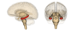
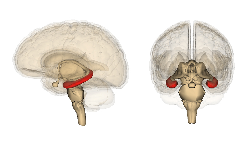
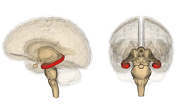
Spatial information from “grid” cells and nonspatial information from” object” cells are combined in the hippocampus (red area), which allows experience to be stored as a long-term memory. Credit: Wikimedia Full size image
|
Mapping Memories
For years, neuroscientists have theorized that memories are like pushpins in a mental map of one’s surroundings. In order for a short-term memory to become a long-term one, according to the theory, it must be coupled with the brain’s representation of the location where the event occurred. It is thought that this coupling occurs in the hippocampus region of the brain, but details of the process remain unknown.
“There are two inputs to the hippocampus; a spatial and a non-spatial pathway,” says James Knierim of Johns Hopkins University, a visiting investigator at the MBL and co-director of the MBL Neural Systems & Behavior course. “We think the hippocampus puts the two together to allow for memory creation.” While the spatial pathway contributes to a cognitive map of the environment, the non-spatial pathway transmits information about items and experiences within the environment, without tracking their location.
Using the rat as a model system, Knierim is investigating certain neurons in the non-spatial pathway that are active when objects in the organism’s environment are present, but hardly fire at all when the objects are taken away.
“We don’t know what it is about these objects that causes these cells to fire. Right now, we just know that they do,” Knierim says. “Do the cells fire regardless of the type of object? Does it make a difference if the object is just giving the rat information about where it is? What happens if we move the object around the environment—do the cells retain memory for where it used to be?”
Preliminary evidence shows that the cells do indeed retain memory of an object’s previous position. The results indicate that information about the object may have been plotted onto a cognitive map as a result of hippocampal processing.
“It’s a very interconnected, complex network of brain areas, and we’re just scratching the surface now, trying to get a handle on how it works,” Knierim says.
Knierim believes the results of this study will be directly applicable to humans, even if the details are not identical. In fact, he says, there is some evidence that the spatial input pathway to the human hippocampus uses the same kind of cells—grid cells— as the rat pathway.
If the firing of a grid cell is plotted on a map of a rat’s environment, the firing points occur at very regular intervals, forming a grid-like pattern. “That turns out to be the major spatial input to the hippocampus. It’s the ‘graph paper’ that sets the coordinates of cognitive spatial representation.”
The interactions between grid cells, “object” cells, and other cells involved in memory formation are complex, Knierim says. “Neuroscience is heading toward no longer concentrating on one brain area at a time but looking at the interactions between brain areas. Brain regions all have to work in a coordinated fashion to produce a final behavior or to produce a perception,” he says.
Knierim’s research at the MBL is funded by the Charles Evans Foundation and the M.G.F. Fuortes Fellowship Fund.
|
 |
|
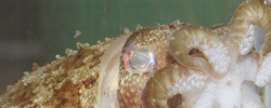
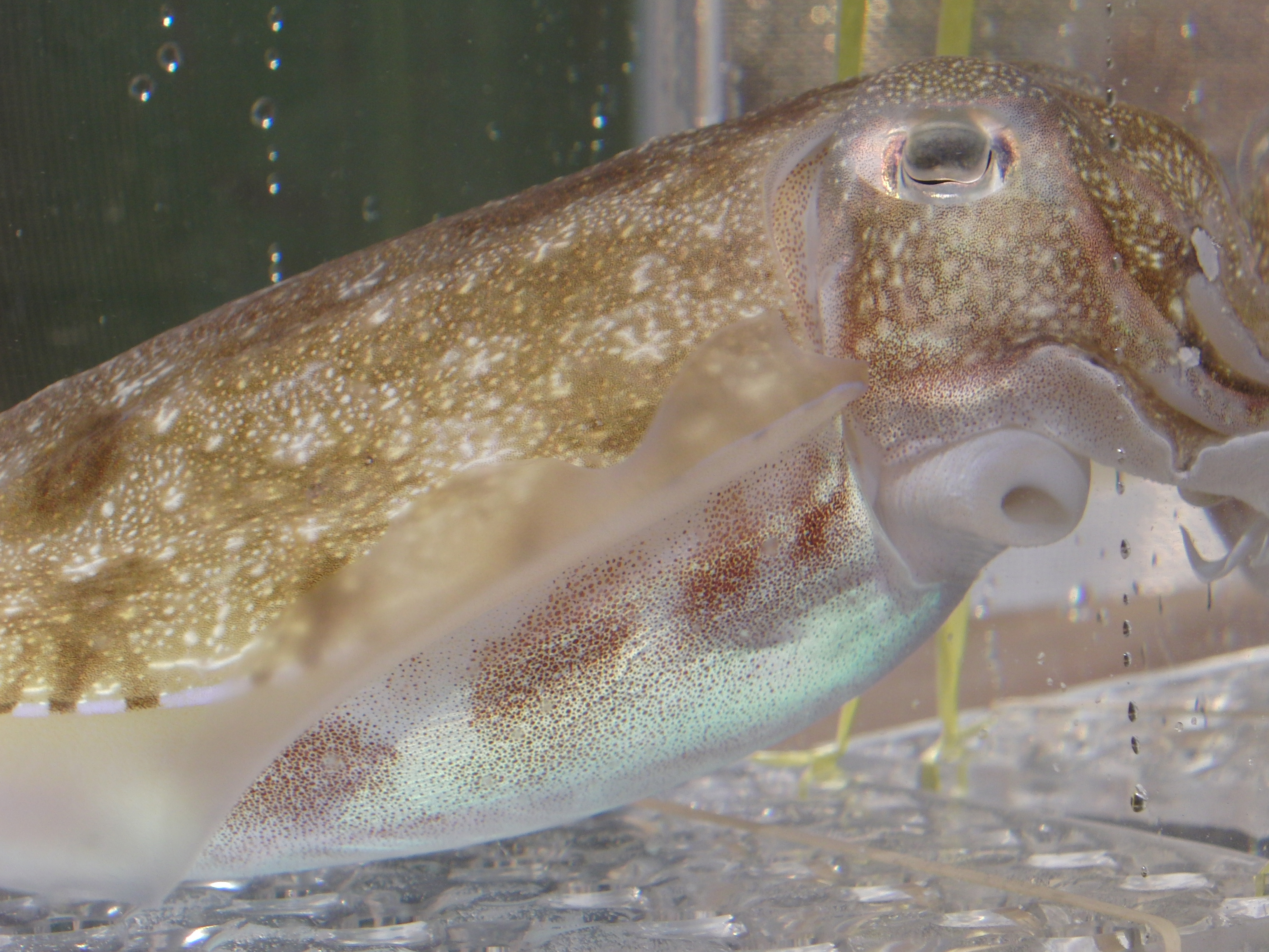
A cuttlefish expresses bumpy skin papillae. Credit: Melissa M. Coates. Full size image
|
|
Blending In
Proteus, the form-changing sea god of Greek myth, must have learned a thing or two from the cuttlefish. In less than a second, the creature can disappear into its surroundings by changing both the color and texture of its skin.
The cuttlefish is a member of the cephalopods, a group of marine invertebrates that also includes squid and octopus. While color changes in cephalopod camouflage have been extensively explored, very little is known about how the animals control three-dimensional skin texture changes.
Melissa M. Coates, an MBL Grass Fellow and a post-doctoral researcher at UCLA, is studying how cuttlefish control expression of skin papillae, bumps on the skin that are raised or lowered to alter texture.
“They’re obviously assessing something about the visual environment and deciding whether or not to express these skin papillae,” Coates says. “The goal is to look for nervous control of these skin papillae and how that’s working.”
To address the problem, Coates has teamed up with several other scientists at the MBL this summer, including Justine Allen, a student in the Brown-MBL Graduate Program in Biological and Environmental Sciences; Kelly Harrington, an undergraduate at Bridgewater State College; and MBL Senior Scientist Roger Hanlon.
|
 |
|
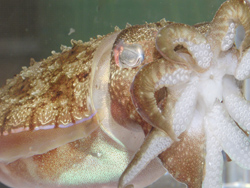
Here, a cuttlefish shows very little expression of skin papillae, taking on a smooth texture. Credit: Melissa M. Coates. Full size image
|
Coates has begun her project by stimulating nerve cells associated with skin papillae in the cuttlefish Sepia pharaonis. This experiment will allow her to characterize nervous system activity during changes in skin texture. She also plans to stimulate various regions of the brain to see how they affect papillae expression. Ultimately, she will test how skin papillae are expressed in response to different visual stimuli.
The results of Coates’ work on cuttlefish will also provide insight into skin texture changes in octopus and squid, which are likewise known for their dynamic camouflage talents. “Cephalopods have some of the most amazing camouflage abilities of any animal,” Coates says. “They are so good at camouflage because they have so many different visual predators with different visual systems.”
While Coates is fascinated with cephalopods out of pure scientific curiosity, she also hopes her work will help raise public awareness about the creatures and their marine environment. “It’s so important for people to know about what’s in the oceans and how humans affect the oceans,” she says.
--By Sarah Stanley
|
The MBL is a leading international, independent, nonprofit institution dedicated to discovery and to improving the human condition through creative research and education in the biological, biomedical and environmental sciences. Founded in 1888 as the Marine Biological Laboratory, the MBL is the oldest private marine laboratory in the Western Hemisphere. For more information, visit www.MBL.edu.
|