Home» News» Press Releases» 2010» Way to Go: MBL Scientists Identify Driving Forces in Human Cell Division
For further information, contact the MBL Communications Office at (508) 289-7423 or e-mail us at comm@mbl.edu
For Immediate Release: March 9, 2010 Contact: Diana Kenney, 508-289-7139; dkenney@mbl.edu
Resources
Citation: Jaqaman, K., et al. (2010) Kinetochore alignment within the metaphase plate
is regulated by centromere stiffness and microtubule depolymerases. J. Cell Biol. 188, 665–679.
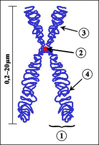
Scheme of a chromosome. (1) Chromatid: one of the two identical parts of the chromosome. (2) Centromere: the point where the two chromatids link. Located at the centromere are the kinetochores, where the microtubules attach. (3) Short arm of chromosome (4) Long arm of chromosome. Credit: Wikimedia.
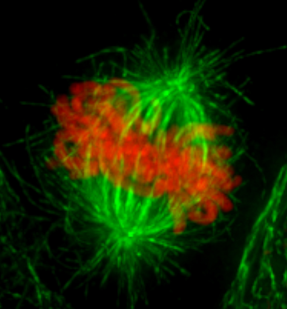
Metaphase in a human cervical carcinoma (HeLa) cell. Chromosomes (red), microtubules (green). Credit: Jason Swedlow, University of Dundee. Full size image.
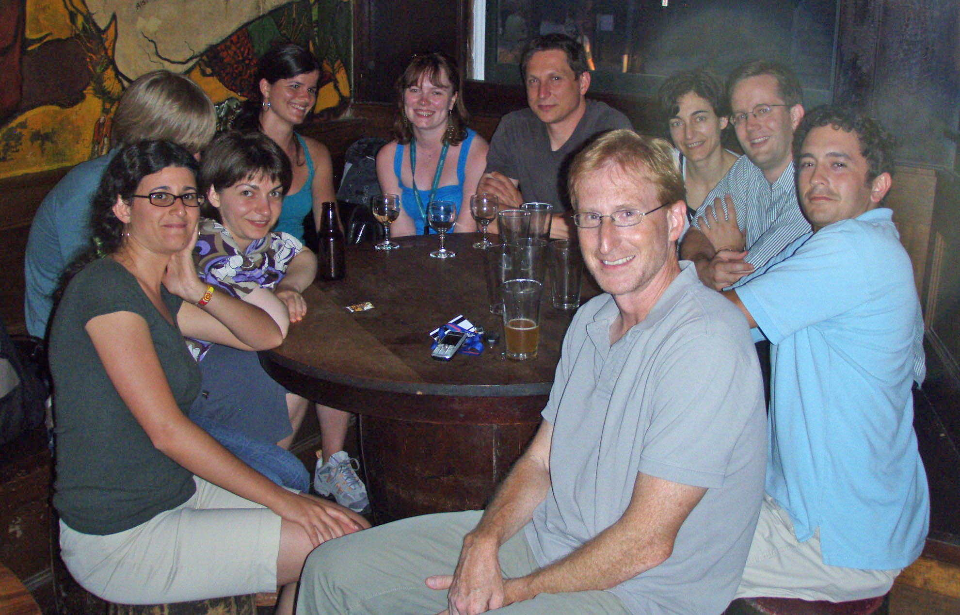
Members of the MBL Kinetochore Consortium in Woods Hole. Credit: Courtesy of MBL. Full size image.
Way to Go: MBL Scientists Identify Driving Forces in Human Cell Division
MBL, WOODS HOLE, MA—If you can imagine identical twin sisters at rest, their breath drawing them subtly together and apart, who somehow latch onto ropes that pull them to opposite sides of the bed—you can imagine what happens to a chromosome in the dividing cell.
Understanding the forces that drive chromosome segregation – a crucial aspect of human development and some diseases, including cancer – is the goal of an international group of researchers who collaborate each summer at the MBL.
In a paper published this week, the group describes newly discovered interactions between sister kinetochores—the protein bundles at the contact point between the two identical strands of a chromosome—and microtubules, the “ropes” that attach to the kinetochores to pull the strands apart.
To do this, the group developed a novel pipeline for preparing and photographing dividing human cells, as well as computational image analysis to quantify the interplay of sister kinetochores in three dimensions.
“We believe we have developed new methods and gained insights that simply aren’t available anywhere else. We couldn’t have done this work anywhere except at the MBL,” says Jason Swedlow, a professor at the University of Dundee in Scotland.
Swedlow’s MBL collaborators on this work, which will continue in future summers, include scientists from the laboratories of Prof. Gaudenz Danuser (Harvard Medical School), Dr. Patrick Meraldi (ETH Zurich, Switzerland), and Dr. Andrew McAinsh (Marie Curie Research Institute, England). The group is known as the MBL Kinetochore Consortium.
—###—
The MBL is a leading international, independent, nonprofit institution dedicated to discovery and to improving the human condition through creative research and education in the biological, biomedical and environmental sciences. Founded in 1888 as the Marine Biological Laboratory, the MBL is the oldest private marine laboratory in the Americas. For more information, visit www.MBL.edu.
The kinetochores are force-generating machines that play an important role in the division of one cell into two. In this time-lapse video, the kinetochores (white dots) of a human cell, which are attached to cell’s chromosomes (not visible), are seen moving to the opposite poles of the dividing cell, where two new daughter cells form. The kinetochores are visible because they have been tagged with a fluorescent protein derived from the jellyfish Aequorea. Video recorded by Ana Amaro of ETH Zurich, a member of the MBL Kinetochore Consortium.