|
For further information, contact the MBL Communications Office at (508) 289-7423 or e-mail us at comm@mbl.edu
For Immediate Release: November 29, 2007
Contact: Diana Kenney, 508-289-7139; dkenney@mbl.edu
The OosightTM Lights the Way: Microscope System Based on MBL Technology Illuminates Stem-Cell Research
MBL, WOODS HOLE, MA — A noninvasive, polarized light microscope invented at the Marine Biological Laboratory (MBL) played a crucial role in a recent breakthrough in embryonic stem-cell research aimed at developing medical therapies...More >>>
Resources
Photos and Video:
Photos 1 and 2 and Figure 1 were published as Supplementary Information with Byrne et. al. (2007) Nature 450: 497-502. Please click on images for full size.
 |
|
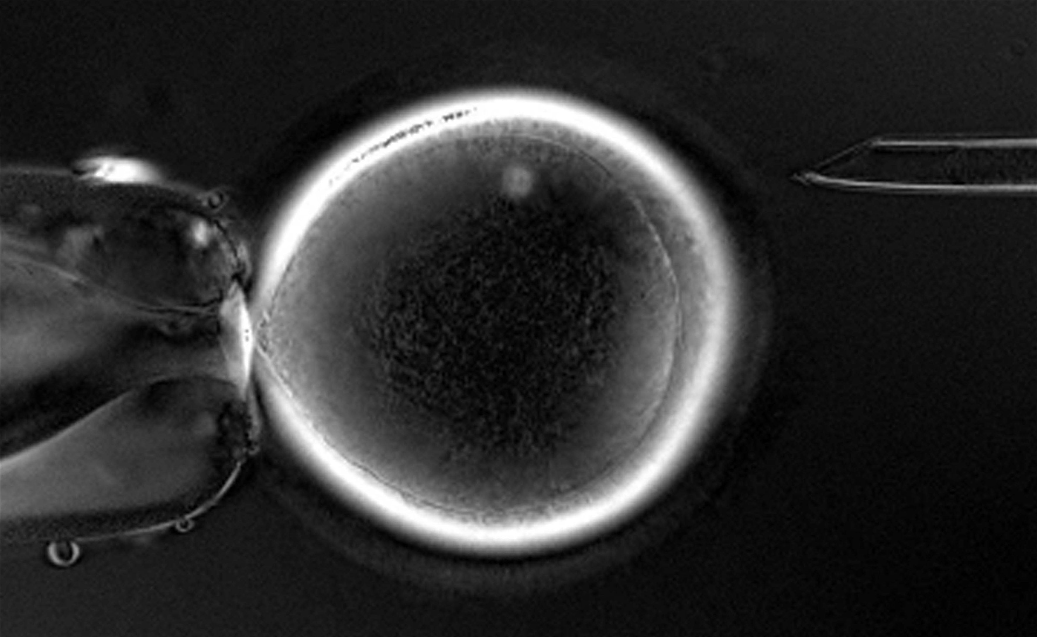
Photo 1 Spindle: Visualization of the meiotic spindle in a rhesus monkey oocyte (egg) using the OosightTM spindle imaging system during enucleation. The spindle is near the 12 o'clock position in the egg.
Image: From Byrne, et al. 2007. Nature 450: 497-502 (Supplementary Material). |
|
|
|
 |
|
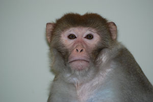
Photo 2 Semos: Semos, the adult rhesus macaque male whose skin cells were used to derive embryonic stem cells.
Image: From Byrne, et al. 2007. Nature 450: 497-502 (Supplementary Material). |
|
|
|
|
|
|
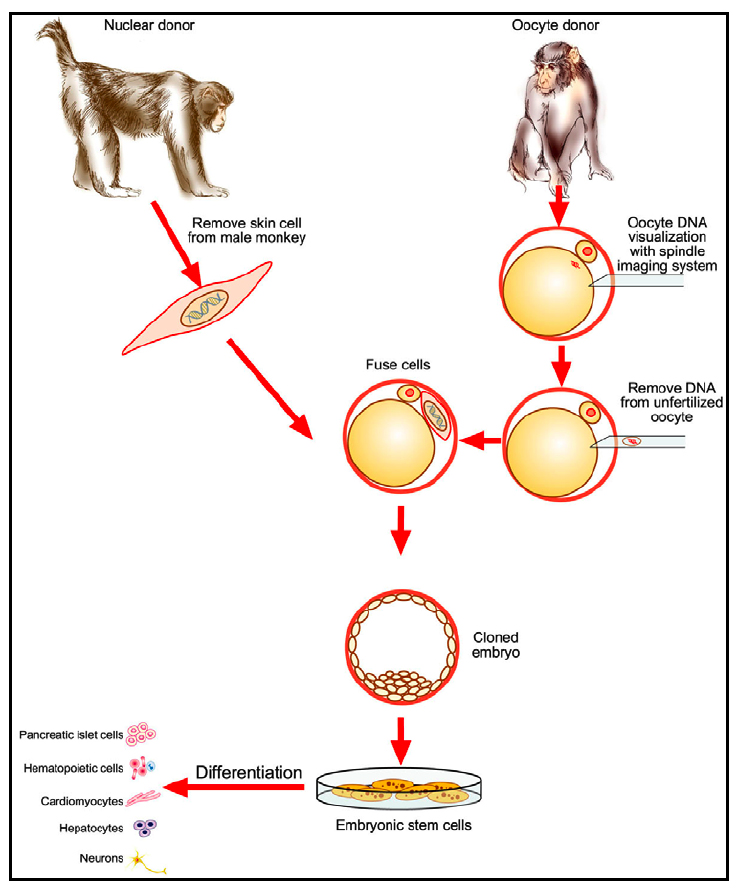
Figure 1 Monkey ESC: A schematic diagram demonstrating the experimental steps involved in the reprogramming of adult primate somatic cells into pluripotent embryonic stem cells. Donor somatic nuclei from skin cells were introduced into enucleated oocytes (eggs) and the resulting embryos gave rise to embryonic stem cell lines that were genetically identical to the nuclear donor male (Semos).
Image: From Byrne, et al. 2007. Nature 450: 497-502 (Supplementary Material). |
A video showing enucleation of a rhesus monkey egg is available for download at:
http://www.cri-inc.com/products/oosight.asp
Citation:
Byrne, J.A., D.A. Pedersen, L.L. Clepper, M. Nelson, W.G. Sanger, S. Gokhale, D.P. Wolf, and S.M. Mitalipov (2007). Producing primate embryonic stem cells by somatic cell nuclear transfer. Nature 450: 497-502.
Mitalipov, S.M., Q. Zhou, J.A. Byrne, W.Z. Ji, R.B. Norgren, and D.P. Wolf. 2007.
Reprogramming following somatic cell nuclear transfer in primates is dependent upon nuclear remodeling. Hum Reprod. 22 (8): 2232-42
Background on OosightTM and LC-PolScope (AbrioTM):
Shribak, M. and R. Oldenbourg. 2003. Techniques for fast and sensitive measurements of two-dimensional birefringence distributions. Appl. Opt. 42: 3009-17.
Liu, L., R. Oldenbourg, J.R. Trimarchi, and D.L. Keefe. 2000. A reliable, noninvasive technique for spindle imaging and enucleation of mammalian oocytes. Nature Biotech. 18: 223-225.
Oldenbourg, R. 1999. Polarized light microscopy of spindles. Methods Cell Biol. 61: 175-208.
Keefe, D., Tran, P., Pellegrini, C., and Oldenbourg, R. 1997. Polarized light microscopy and digital image processing identify a multilaminar structure of the hamster zona pellucida. Human Reprod. 12: 1250-1252.
Oldenbourg, R. 1996. A new view on polarization microscopy. Nature 381: 811-2.
Oldenbourg, R., and Mei, G. 1995. New polarized light microscope with precision universal compensator. J. Microscopy 180: 140-147.
Inoué, S., and H. Ritter, 1975. Dynamics of mitotic spindle organization and function. In S. Inoue and R.E. Stephens, eds., Molecules and Cell Movement. Raven Press, New York, NY, p. 3-30.
Inoué, S., and W.L. Hyde. 1957. Studies on depolarization of light at microscope lens surfaces. II. The simultaneous realization of high resolution and high sensitivity with the polarizing microscope. J. Biophys. Biochem. Cytol. 3: 831-838.
Inoué, S. (1953). Polarization optical studies of the meiotic spindle. I. The demonstration of spindle fibers in living cells. Chromosoma 5: 487-500.
The OosightTM uses the following MBL patents licensed to CRi:
- Oldenbourg, R., and G. Mei. “Polarized light microscopy,” U.S. Patent 5,521,705
- Shribak, M., and R. Oldenbourg. “Retardance measurement system and method,” U.S. Patent 7,202,950
- Shribak, M., and R. Oldenbourg. “Retardance measurement system and method,” U.S. Patent 7,239,388
Links:
|