|
|
|
|
|
|
|
|
|
|
|
 |
|
September 18, 2008
The Power of Self-Healing
MBL Visiting Researcher Explores Wound Repair in the Comb Jelly
By Joseph Caputo
MBL, WOODS HOLE, MA—We’d save a fortune on bandages if we had the regenerative abilities of a comb jelly: Blisters from a night out in uncomfortable shoes would heal in minutes, surgical scars would be a thing of the past, and cuts would heal overnight. How a 500+ million-year-old creature can have this capacity, while humans don’t, is the focus of ongoing investigation at the MBL.
PHOTOS: Click on thumbnails for larger images.
|
|
|
|
|
|
|
|
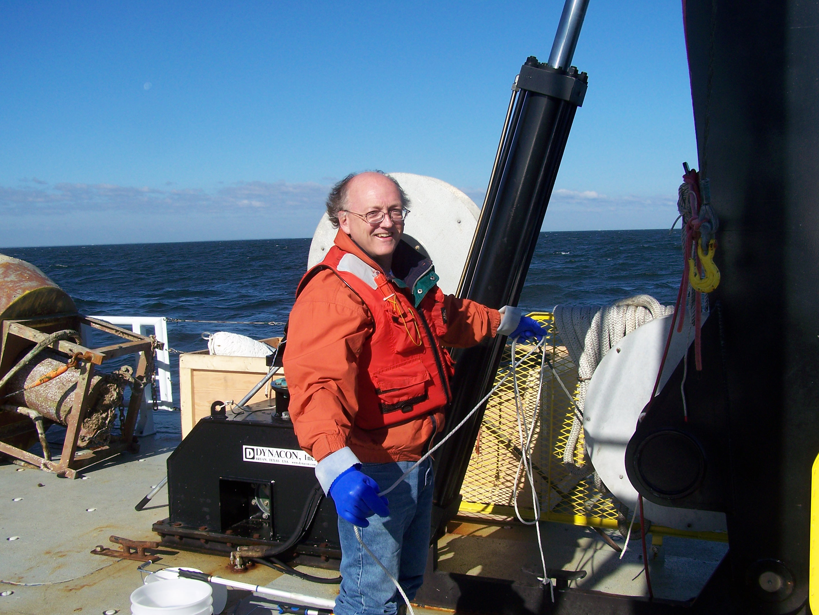
MBL visiting investigator Anthony Moss preparing to catch ctenophores on the Chesapeake Bay on the R/V Hugh Sharp (Credit: Anthony Moss)
|
|
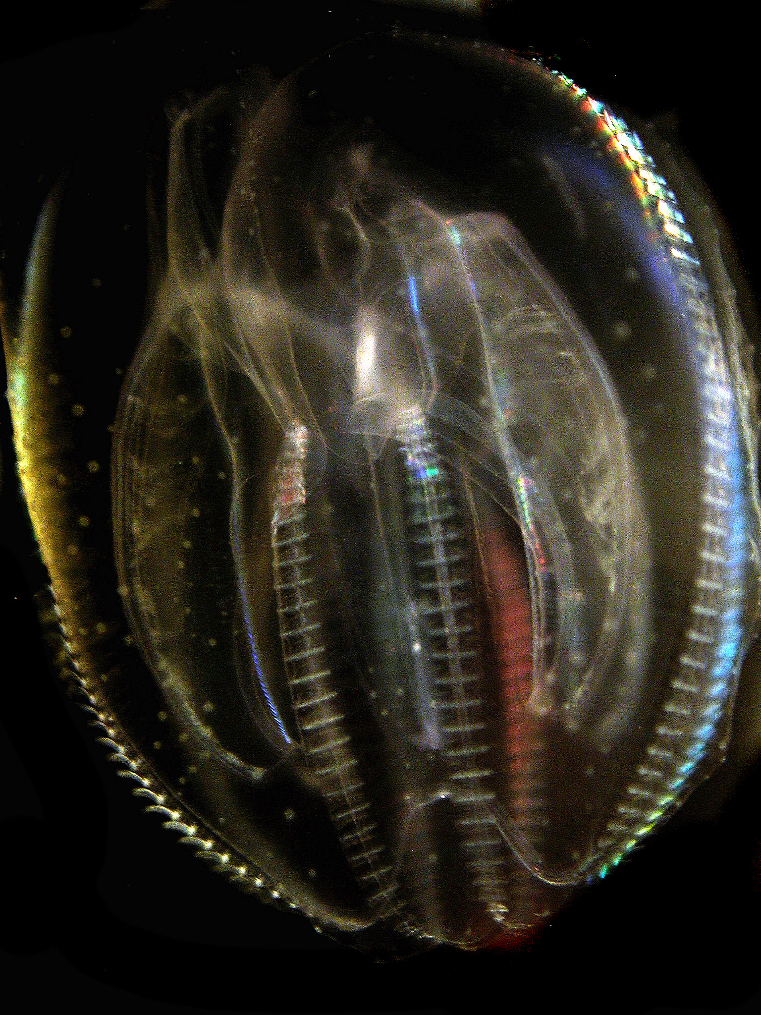
The sea walnut, Mnemiopsis leidyi, can quickly repair itself - a few minutes to a few hours depending on the injury - without scarring. (Credit: Anthony Moss)
|
|
|
|
|
|
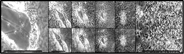
An epithelial wound heals to scar-free completion in a little over an hour. Center upper row shows the surface; lower row focuses 3 mm under the ‘skin.’ All scales: 1 mm.
|
|
To study wound repair is to rummage through a time capsule of evolutionary innovation, which is one of the reasons why Marine Biological Laboratory (MBL) visiting investigator Anthony Moss finds comb jellies so intriguing. Moss has studied the jellies, known scientifically as ctenophores (pronounced TEEN-o-fours), for more than 25 years. He is one of the world’s experts on the ancient animals, having made multiple discoveries about their physiology. Moss is now focusing his attention on their capacity for wound healing and regeneration, which is commonly studied in other regenerative marine organisms, such as starfish.
“Ctenophores are endlessly fascinating,” says Moss, an associate professor of biology at Auburn University in Alabama. “They are excellent model organisms for the study of epithelial cell biology and smooth muscle because those cell types are easily accessed and manipulated in these animals,” he says, referring to the large, smooth muscle fibers and paddle-shaped ctenes, or comb plates, which are made of many thousands of hair-like cilia. (They are much like those in our lungs except that they are very large and on the outside of the animal). Although comb jellies resemble small jellyfish, hence the name, ctenophores bear comb plates and lack stinging cells, or nematocytes, which are found in true jellyfish.
Ctenophores have long interested biologists who are curious about the evolution of nervous systems, the function of smooth muscles, and the structure and function of cilia, work that was pushed forward by the efforts of Moss’s MBL mentor, Sidney Tamm of Boston University. More recently, ctenophores have been closely examined by MBL visiting investigator Mark Martindale of the University of Hawaii, who examines their development and evolution using molecular methods. A recent publication by Martindale and his collaborators suggests that ctenophores are one of the most ancient organisms on the planet.
Comb jellies may also prove to be an excellent model organism for wound repair and regeneration research. Moss is at the MBL this summer to investigate how the comb jelly called the sea walnut, Mnemiopsis leidyi, can quickly repair its skin-like epithelium in a few minutes to a few hours – depending on the injury – without scarring. Using high resolution differential interference and fluorescence microscopy, Moss wants to describe the wound healing process at the cellular and molecular level. At the moment, he believes it is the recruitment and multiplication of many thousands of cells located in M. leidyi’s mesoglea, or jelly, to the wound site, that is responsible.
“The wound-healing cells crawl rapidly to the edge of the wound site, and bind tightly to each other,” says Moss. “Cells then produce long processes that reach out across the void created by the cut and attach to cells at other locations around the wound edge. The newly-recruited cells contract and pull against each other, steadily pulling the edges of the wound closed. They also bind to smooth muscle cells that underlie the epithelium, thereby stabilizing the wound repair complex. They act much like a purse-string: thousands of cells migrate to a wound and by pulling against each other, rapidly shrink it to a miniscule size. New cells arrive and using the existing cells as a scaffold, quickly climb on top of each other, and contract in a cooperative manner to seal the wound.” Very likely, this represents one of the most ancient mechanisms for healing a wound: simply close it up.
According to Moss, rapid wound repair is critical to ctenophore survival. The ocean is packed with merciless microbes and even the smallest open wound could mean a deadly infection. A ctenophore is mostly water surrounded by a very thin but flexible and rubbery epithelium. Because they have so little total tissue mass, a small cut on its epithelium could cause easily result in all of its internal fluids to “bleed” or leak out. This is problem as the ctenophore’s delicate skin is regularly nicked by rocky shores or the sharp edges of eelgrass and preyed upon by small fish or shrimp that unceasingly nip at the jelly. In a healthy ctenophore, probably hundreds of small wounds are quickly and simultaneously healing, and in this manner, the ctenophore remains ahead of the microbes. Even after having had as much as 50% of the body bitten away, the ctenophore can close the huge wound, and regenerate a normal body in just a few days.
“In this first stage of the work, we are setting out to describe just what happens at the cellular level when the ctenophore heals a wound,” Moss says. “However, with my master’s student Matt Dodson, I am also moving on to the second stage: to find the molecular players that correlate with and could drive the wound healing response. “From that point, I hope to learn which molecules involved in ctenophore wound repair could be useful for a better understanding of human medical care delivery.”
“The fantastic microscopy facilities here at the MBL were a major reason why I wanted to come this summer. There are basically no other research stations that combine the easy access to the animals with the excellent laboratory facilities in the MBL Central Microscopy Facility. And of course, the opportunity to learn from the world’s finest scientists affords new insights into my work,” Moss says.
Moss’s research begins at an exciting time in MBL’s history, as it begins the initial stages of creating a Center for Regenerative Biology and Medicine. The research initiative aims to establish the MBL as a leader in the field of regenerative and stem cell biology.
|
| |
|
|
|
|
|