 |
 |
|
 |
 |
 |
 |
 |
|
 |
 |
 |
 |
Fasten Your (Nano) Seatbelts!
 |
|
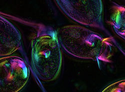
Live Vorticella visualized in the LCPolScope. Intensity encodes retardance and color encodes direction of fiber slow axis.
|
|
A Centrifuge Polarizing Microscope (CPM) invented by MBL scientist Shinya Inoué, recently enabled biological engineer Danielle Cook France to study the force, speed, and fuel source of a tiny spring that is, gram for gram, the most powerful known engine in biology. The spring drives the microscopic protozoan, Vorticella, and the new findings could rev up the creation of nano devices.
France, a graduate student in biological engineering from MIT who worked with scientists and microscope inventors in the MBL's Architectural Dynamics in Living Cells Program to look under the hood of the unicellular Vorticella, presented her research at the 45th meeting of the American Society of Cell Biology.
The Vorticella is a bell-shaped aquatic organism that attaches to submerged rocks or plants with a contractile stalk that recoils rapidly when the creature is disturbed. Scientists have long known that relative to its size the spasmoneme, the spring that lies inside and powers the stalk, is extremely forceful. France and her colleagues observed the spring as it pulled against the centrifugal force of the CPM and found it to be more powerful than previously thought—weight for weight even more powerful than a car engine.
|
|
|
Contracted (in 10 mM dibucaine) Vorticella stalk visualized in the LCPolScope.
|
The research also sheds new light on how calcium ions fuel this nanospring. The MIT scientists identified a specific protein, centrin 5, that responds to the calcium signal. According to France, this is one of the first concrete links between the fuel source, the protein components, and the resulting movements.
"This leads us to a closer understanding of two things: how cells use centrin-based engines to generate enormous forces and how we can possibly reconstruct centrin-based materials and devices for our own use at the micrometer and nanometer scales," says France.
Still images captured using the LCPolScope, developed by Rudolf Oldenbourg, Guang Mei, and Mykhailo Shribak with assistance from Grant Harris at the Marine Biological Laboratory, Woods Hole, MA. Video captured using the Centrifuge Polarizing Microscope, developed by Shinya Inoué at the Marine Biological Laboratory. Images by Danielle France.
Additional high-resolution photos
Movies
Live Vorticella contracting in real time. Image courtesy of Arpita Upadhyaya and Alexander van Oudenaarden, MIT
Live Vorticella cell contracting and re-extending under low opposing centrifugal force (~21g acceleration) in the Centrifuge Polarization Microscope.
Live Vorticella cell contracting and re-extending under high opposing centrifugal force (~11500g acceleration) in the Centrifuge Polarization Microscope.
|
| |

 |
|
 |
 |
|