
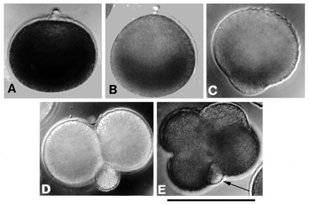
A. Formation of the second polar body. The egg is slightly flattened at the animal pole, where the polar bodies are forming.
B. Early mitosis in which the egg is now elongated at the animal pole into a "pear shape."
C. Late mitosis showing the beginning of the formation of the polar lobe at the vegetal pole.
D. Cytokinesis of the first cell division. The polar lobe has reached its final size and is ready to be resorbed into the CD cell.
E. Cytokinesis of the second cell division. A Second polar lobe (arrow) has formed in association with the D cell.
Scale bar = 100 µm. From Eckberg and Anderson (1995).