|
|
|
|
|
|
|
|
|
|
|
 |
|
|
| |
This story appeared in:
MBL Monthly, February 2003, Vol. 12, No.2 | Return to Table of Contents
LabNotes, Summer 2003, Vol. 13, No.2 | Return to Table of Contents
Pol-Scope Allows Scientists to See Cells in a New Light
If you look at a living cell under a light microscope, chances are you don't see much. Most of the structures inside a cell are transparent to ordinary light, making them indistinguishable from their aqueous background. In order to visualize the internal structure of cells, scientists have had to settle for other methods of creating contrast, such as staining and fluorescent labeling, that typically only allow them to study the cells after they are dead.
But Dr. Rudolf Oldenbourg, a Senior Scientist at the Marine Biological Laboratory, has developed a microscope that enables investigators to view cells while they are alive and in action. As part of the Architectural Dynamics in Living Cells Program at the MBL, Dr. Oldenbourg's lab focuses on developing optical technology to image and manipulate living cells. "Specimen preparation is a big factor in the success of what you are looking for," he says. "It is an art as much as a science." By taking advantage of a natural property of cellular materials to "create contrast where there was no contrast" before, Oldenbourg's "Pol-Scope" allows scientists to study cellular processes as they occur.
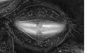
The Pol-Scope uses polarized light to analyze the birefringence of a specimen. As light waves travel, they oscillate in any direction perpendicular to the direction in which they are traveling—a light wave moving toward you might be vibrating up and down, side to side, in ellipses or circles. In ordinary or scattered light, this direction of oscillation changes so rapidly that the waves appear to vibrate in every direction. Polarized light, however, consists of waves that are oscillating in only one direction, such as up and down.
Birefringence is a property of certain materials in which the index of refraction, or the degree to which the path of light is bent as it passes from a material of one density to another, differs between perpendicular planes of polarized light. For example, polarized light that is oscillating up and down may be bent more when it passes through a birefringent material than polarized light that is oscillating from side to side. Many biological structures, such as the mitotic spindle or membrane stacks, exhibit molecular order and are therefore birefringent. Because different structures exhibit different orientations and degrees of birefringence, this property can be used to distinguish many of the structures within a cell.
Although polarized light microscopes have been around for a while, the Pol-Scope features several improvements over the traditional models that make it much easier to use. When using a traditional polarized light microscope the specimen has to be oriented correctly with respect to the fixed polarization axis of the microscope. Different structures in a living cell usually have different orientations and therefore the entire specimen had to be rotated for observing different structures, making the microscope very cumbersome. The Pol-Scope, however, uses a device called a universal compensator that automatically changes the plane of polarization relative to the specimen by altering the voltage that is applied to a pair of optical modulators. With this device, the Pol-Scope is able to simultaneously measure the birefringence in every resolved specimen point, across the entire viewing field of the microscope; traditional models could only analyze a single image point at a time, so they were very time-consuming to use. The Pol-Scope uses an electronic camera to rapidly take four images of the specimen that is illuminated with different orientations of polarized light. These images are analyzed and combined with algorithms programmed into an ordinary desktop computer to produce a detailed picture of the birefringent structures within the cell in less than a second.
The relative ease-of-use and efficiency of the Pol-Scope have made it much more practical for applications in biological research. Oldenbourg's lab is now collaborating with several investigators at the MBL to use the Pol-Scope for studying a variety of cellular processes, including actin-based and tubulin-based systems that are involved in cell division and cell motility.
A commercial version of the Pol-Scope, called the LC-PolScope, has been developed by Cambridge Research and Instrumentation in Woburn, MA, which licensed the patented Pol-Scope technology. Oldenbourg is now working with Dr. Michael Shribak on another patent to upgrade the Pol-Scope. This upgrade will enable the microscope to analyze specimen birefringence in three dimensions rather than in a single plane of focus.
— Angela Damery
|
|
|
|
|