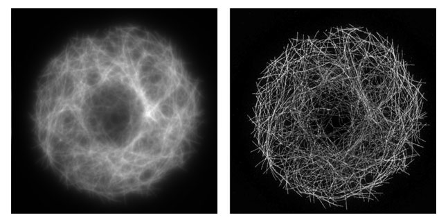
Comparison of super-resolution and conventional images of microtubules in a Drosophila S2 cell, collected as part of the MBL's Neurobiology course in Summer 2011. At left, a diffraction limited image obtained with conventional fluorescence microscopy. At right, a structured illumination image (SIM). Note the two-fold increase in spatial resolution in the SIM image compared to the diffraction limited image. Images were collected on a Zeiss Elyra Super-resolution microscope by Chris Rieken. Images are available from The Cell: An Image Library ( www.cellimagelibrary.org ). The z-series are available in the Cell Image Library as CIL 36797.
|
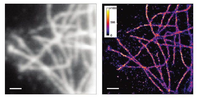
Comparison of super-resolution and conventional images of fixed microtubules by Paul Selvin. Left: Normal fluorescence image. Right: Generalized single molecule high-resolution imaging with photobleaching (gSHRImP) image. From: Simonson, P.D., Rothernberg, E., and Selvin, P.R. (2011) Single-Molecule-Based Super-Resolution Images in the Presence of Multiple Fluorophores. Nano Lett.:11, 5090–5096.
|