
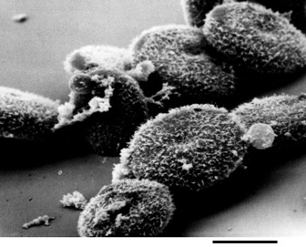
The flattened disc shaped cells possess surface microvilli, and have a slight depression in the nuclear area. SEM; bar = 40µm.
|
|
| Figure 6. Scanning electron micrograph of freshly shed coelomic oocytes prior to GVBD.
The flattened disc shaped cells possess surface microvilli, and have a slight depression in the nuclear area. SEM; bar = 40µm. |