
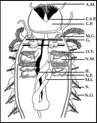
The dissection is cut in the midline and spread laterally, showing position of the ovaries and attached nephromixia. For greater detail of nephromixium and attached ovary, see Fig. 4.
AM - antennular membraneCSP - cephalic spinesCP - cephalic plaqueMG - mucus glandG - gillOV- ovaryNM - nephromixiumH - heartNP - nephridioporeMI - middle intestineS - setaeNO - notopodium
(From Tweedell, 1966)