
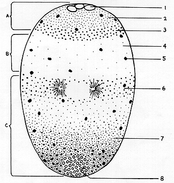
The stratified particles were stained by different vital dyes, fluorochromes and cytochemical tests. The three zones of the unstained centrifuged egg (A, B and C) are indicated, beginning with zone A at the centripetal pole.
- Fat droplets
- Lipid granular cap
- Granular band I
- Hyaline zone with diffuse granules
- Cortical granules
- Granular band II, mitochondria and protein yolk
- Dense protein yolk
- Centrifugal vacuole with basophilic granules
(From Tweedell, 1962).