
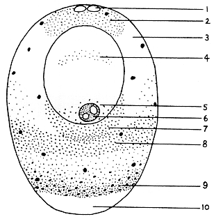
- Fat droplets
- Lipid granular cap
- Hyaline zone
- Germinal vesicle substance
- Basophilia
- Amphinucleolus
- Granular Band I
- Granular Band II, mitochondria and proteid yolk
- Dense proteid yolk
- Centrifugal vacuole
|
|
Figure 12. Diagram of a centrifuged immature oocyte, showing particle distribution in relation to the intact germinal vesicle
|